It was Hippocrates (460-375 BC) who first observed “should one part of the body be colder or hotter than the other, disease is present in that part”.
With the development of low-cost, highly sensitive, Infrared (IR) cameras, the ability to monitor temperature anomalies has become very easy and provides a valuable sixth sense, especially for those charged with the welfare and care of animals who cannot speak about their injury or infection!
Thermal imaging, or Infrared Thermology (IRT) is a highly intuitive, efficient, contactless temperature measurement technology. It has been found to be an extremely useful diagnostic tool in the detection of infections where the primary indicator is inflammation (characterised by heat and swelling), and thus has become widely applied to animal production, aiding farmers, vets, and other professionals in the healthcare of livestock.
Infrared cameras are more frequently being used in the diagnosis of several economically significant diseases of livestock e.g., mastitis, lameness, bluetongue virus in sheep, and bumblefoot in poultry, and can often detect sub-clinical problems before they become apparent to the naked eye.
Thermal imaging allows you to monitor cattle health without touching or catching the animal, reducing animal stress while maintaining a safe distance. It aids in the early detection of disease and inflammation, promoting cattle health and maximises productivity. Early detection of hoof and udder issues can reduce treatment costs, speed up recovery, and reduce yield losses
Lameness is one of the most debilitating diseases in dairy cows causing pain and affecting animal welfare. It also has a negative effect on reproduction, and reduces milk production.
Healthy cows are productive cows, yet various studies suggest that on average some 20% to 30% of a dairy cow herd are lame. Typical losses due to lameness are estimated to be in the region of £300 per cow (including vet fees but not culling). In a 400-cow dairy herd with 30% lameness that’s an annual loss of around £36,000 to the farmer.
Quantifying losses caused by premature culling, treatment costs, and milk loss due to lameness is important to persuade the dairy farm ecosystem to adopt new technologies such as thermal imaging, to prevent or minimise lameness.
Detection of lameness is most difficult in its early stages. Various existing methods and tools exist for this including observation of animal gait, either manually or by use of accelerometer systems, or by direct observations during hoof care.
One disadvantage of observation techniques can be the subjective views and inconsistencies of the observers, who require an “experienced eye”. Observation is time-consuming, and too often there are lesions on the limb without any visual evidence of lameness. Thermal imaging thus provides observers with a “sixth sense”.


Thermal imaging offers several advantages over traditional diagnosis:
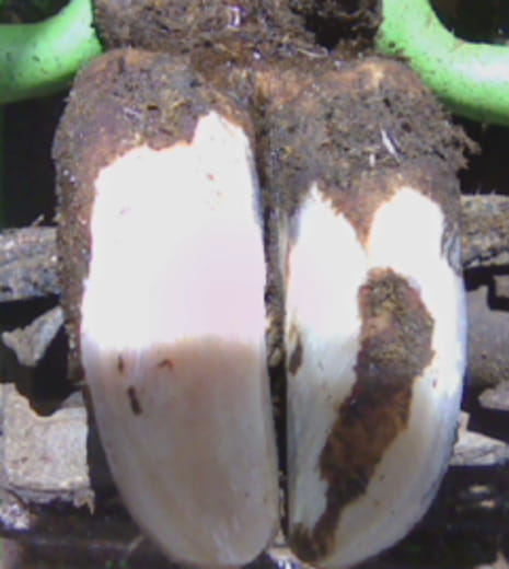
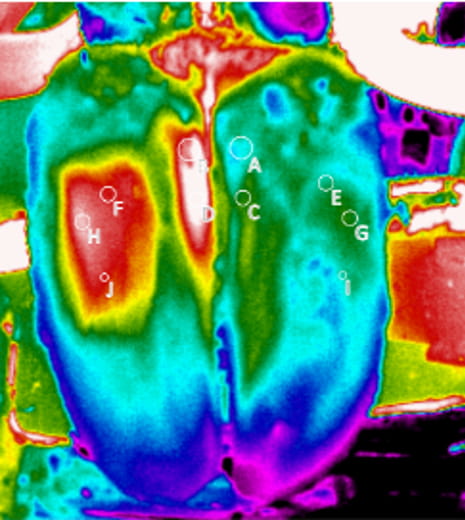
The intuitive nature of thermal imaging aids in the exact localisation of areas of increased (or decreased) temperature, which may indicate inflammation, or decreased blood flow. This allows early detection of lesions on the extremities before the onset of lameness, possibly even before visible symptoms appear, allowing treatment of lame cattle to start earlier.
In a report in Farmers Weekly of March 2018, farmer George Coles says of using thermal cameras – “Straight away you could find that if you had a lame cow, you could see where the hotspots were”. “The hotspots were often higher up in the leg rather than the foot, so you could treat it with anti-inflammatories, not antibiotics.”
On his farm, antibiotics are only used when thermal imaging shows that lameness is caused by inflammation in the hoof area.

Modern thermal cameras are able to detect skin temperature differences of ±0.0 °C with an accuracy of better than ±2 °C, but pragmatically we are typically looking for regions of thermal anomalies and areas where there is a clear contralateral temperature difference. This approach makes the need for “absolute temperatures” less important, and in practice any thermal anomalies are immediately and intuitively obvious.
While it is true that higher resolution cameras allow thermographers to work at a much greater distance from a target without loss of absolute temperature accuracy (due to optical Field of View issues outside the scope of this discussion), some camera manufacturers hold the view that a 1.3cm x 1.3cm area can be accurately measured for temperature by a 320×240 pixel IR imager at a distance of around 18m.
Therefore realistically, in clinical imaging, a high-quality 384×288 pixel IR camera, such as used by Thermidas can be considered sufficiently accurate for most clinical purposes, and indeed, in most cases, animals are imaged from a distance of significantly less than 18 meters.
Thermidas’ own guidelines for thermal imaging of cattle include imaging in a bright location without exposing the subject to direct sunlight, sources of heat, or convective air movements. We also suggest the whole animal should be imaged from around 3m, while for specific areas of interest (e.g., a hoof), a distance of 1 m – 1.5m is ideal.
Byrne et al in a study in Ireland in 2017, indicated that IR measurements made using standard operating procedures can achieve high levels of precision in an agricultural environment, with the usual caveats of following preparation guidelines, and eliminating external sources of radiant heat and convective air.
Another study, this time in Northern Ireland (Seeley at al 2018), also indicate low levels of variability and high levels of repeatability with IR temperature measurements.
Both Byrne and Scoley found that the provision of basic, standardised, IR protocols was sufficient to limit between operator variation with camera operators of varying abilities.
Kriz et al (2021}, in a study in The Czech Republic, stated that “Based on research of scientific articles dealing with IRT in animal production, the claim that IRT allows detecting problem areas noninvasively with a high level of reliability can be made”.
All these findings concur with Thermidas’ own pragmatic experience, that thermal imaging can give reliable, sensitive, quantitative, data, with high levels of repeatability.
Reports from both Warwickshire and Northern Ireland, show that thermal imagers combined with other lameness monitoring systems (such as CowAlert) significantly enhance detection of hoof lameness problems, even when cows show no visible signs of lameness.
In a Warwickshire workshop conducted in May 2018 by Data Driven Dairy Decision For Farmers (4D4F), Innovation for Agriculture’s Richard Lloyd was “astounded” how two different technologies could combine so well.
“Lameness is one of the biggest costs in dairy farming and is often underestimated. I have seen today that thermal imaging cameras, in conjunction with CowAlert’s lameness monitoring, can have a big impact on farm profitability and animal welfare. Cows that showed no visual sign of lameness were identified as lame by technology and treated much earlier, and more accurately, than was possible before.
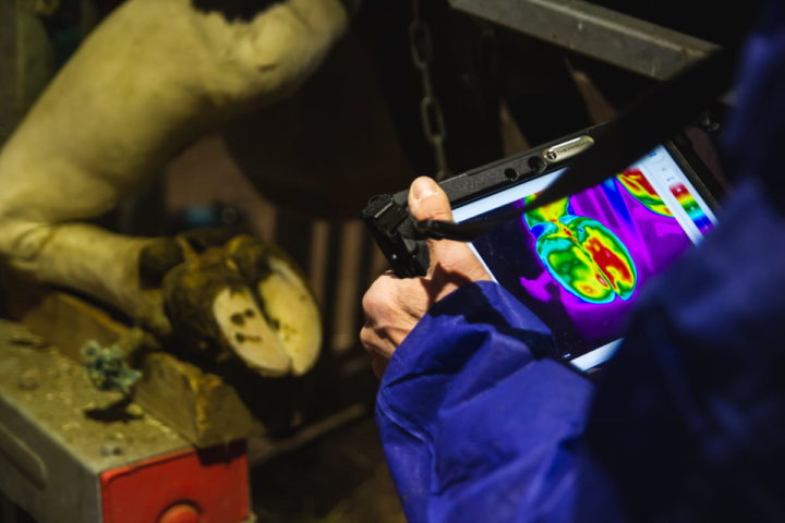
“It should be noted that Thermidas IR system utilises a ruggedised 10 inch Android tablet, and therefore is likely to be compatible with most industry standard farm management and lameness detection software.”
The use of thermal imaging to aid farmers, cattle foot trimmers, and vets in the detection of a variety of diseases in livestock, including mastitis and lameness in cattle, has been shown to be clinically effective, and based on such evidence, should, in time, become a mainstream diagnostic tool. And with a relatively modest, one-off investment in a thermal imaging solution, the average farm can benefit from 10’s of thousands of pounds in annual savings.
Discover our new, portable solution. Compact and easy to use, the IRT-384 Tablet allows you to conveniently take and analyze thermal images on a single device - wherever you go.
Learn more and get yours
Thermidas VET
Polttimonkatu 4
33210 Tampere, Finland