The thermal pattern on the skin of a healthy animal is always symmetrical; there are no differences from side to side. In thermal image analysis, asymmetries that meet the diagnostic criteria are examined in order to locate possible issues quickly and reliably. On its own, thermography may not be enough for reaching a conclusive diagnosis. However, it may critically speed up the detection of injuries and inflammations thus leading to earlier and more accurate treatment.
Thermography reveals even the tiniest abnormalities. Early detection means quicker treatment, which in turn often translates into lower treatment costs and shorter recovery. Thermal imaging is a superb tool for early diagnostics, because it may enable the detection of possible problems even weeks before any clinical signs appear.
Thermal imaging can be used to quickly and reliably detect changes in muscular, vascular, skeletal, and nervous systems.
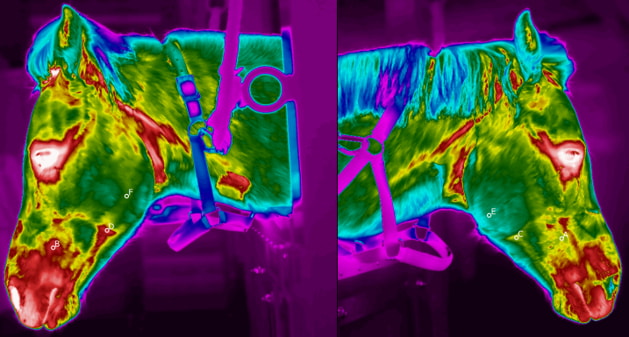
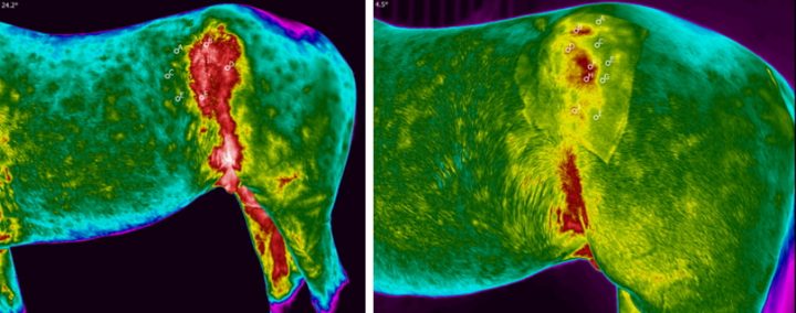
Case 1: Jaw abscess on the left side
A pony stopped eating with no obvious cause. The owner has no clue as the pony was recently purchased and the whole history was not known.
Three different veterinarians had tried to reach a diagnosis with conventional methods but were all unable to conclude what was wrong.
Diagnostic thermal imaging helps to locate an abscess that was causing the problem. The abscess was surgically removed and the pony started to heal rapidly.
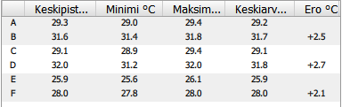
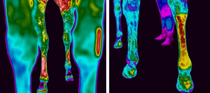
Case 2: Early detection of injuries
A thermal imaging finding on a trotter in the hoof of the right front leg after a hit-out. In an X-ray imaging, changes in the hoof joint and articular capsule full of liquid were found.
Case 3: Monitoring the effectiveness of treatment and progress of recovery
A 6-year-old trotter. Diagnosis: The left iliac crest and the tendon structure surrounding it were accidentally damaged. The area was treated using PRP technology. Follow-up photos every 2 weeks.
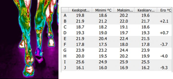
Case 4: Suspensory ligament
A 4-year-old trotter. Diagnosis: injured suspensory ligament (50%) in the right hind leg. Diagnostic thermal imaging is used to monitor treatment impact and the healing process.
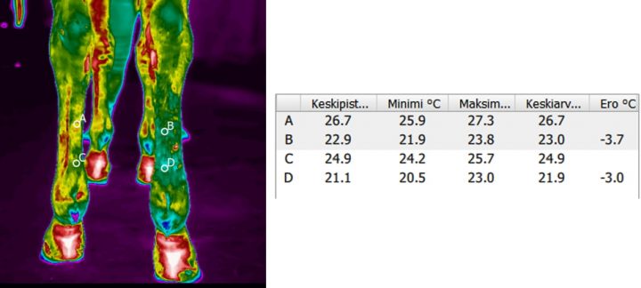
Thermal images of the hind legs before the first PRP-treatment, few days after the injury.
Hind legs from behind 1,5 months after the injury and rehabilitation / equine hydrotherapy. Right hind leg is colder than the left one, because the horse is not putting weight on the injured leg.
After 20 minutes of exercise (water treadmill) the right hind leg has increased circulation, and temperature differences between the left and the right hind leg are closer to normal.
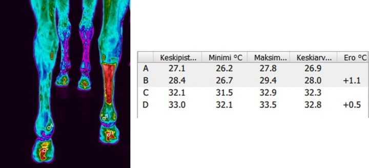
Hind legs 8,5 months after the injury and active rehabilitation.
The right hind leg is still colder than the left hind leg, indicating that the suspensory ligament injury is not completely healed even after 8 ½ months. However the activity level of the horse is now gradually increased and progress is closely monitored.