In August, the horse was diagnosed with hindlimb osteoarthritis, which was treated with injections for two months. In October, the horse had a bad fall in a startle during a cross-country race. Between November and December, his movement had become lame and unsteady. The vet examined the horse from top to bottom and treatment was directed to the hind leg.
Findings
There was no clear response to the treatment of the hindquarters and the owner was already ready to send the horse to greener pastures. The owner contacted equine professional Mari-Tiina Jääskeläinen and the horse was thermographed. Based on the thermal images, the cause of the pain was found to be an inflamed SI joint. The horse has been treated with precision therapy and the pain has been reversed.
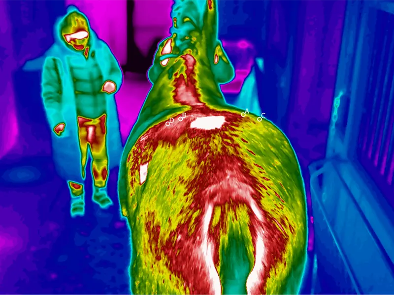
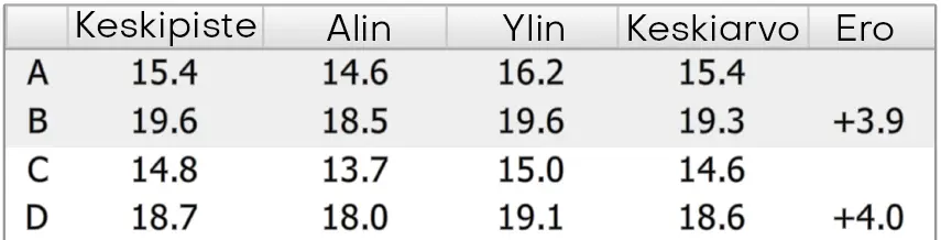
Back from behind. Thermal imaging findings of the lumbar region (B,D).
This injury is often chronic, easily relapsed and difficult to treat. It is often found in sport horses, where a rapid recovery back to working condition is important to the owner. Early diagnosis of the injury is important, so that treatments can achieve better results.
In this client’s case, thermal imaging made the invisible visible and enabled timely treatment. The SI problem was properly treated and the horse won the regional championship for young horses in his region.
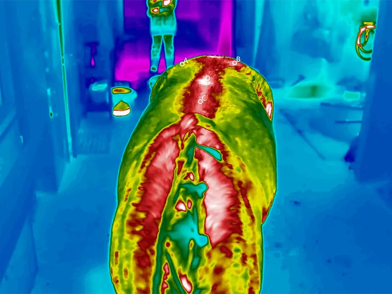
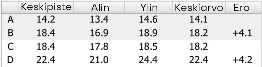
Back to front. Thermal imaging findings of temperature differences in the lumbar (B) and thoracic (D) regions.
IRT-384 Tablet
Discover our new, portable solution. Compact and easy to use, the IRT-384 Tablet allows you to conveniently take and analyze thermal images on a single device - wherever you go.