Horses tend to mask their pain and some injuries may go unnoticed. In this client’s case, the horse’s owner reacted quickly to a change in the horse’s behaviour, which had suddenly become frightened and nervous.
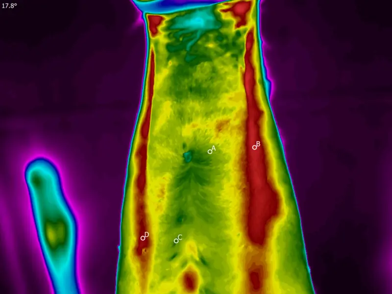
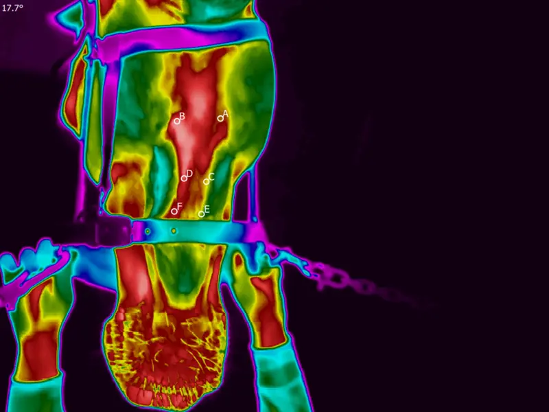
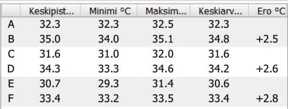
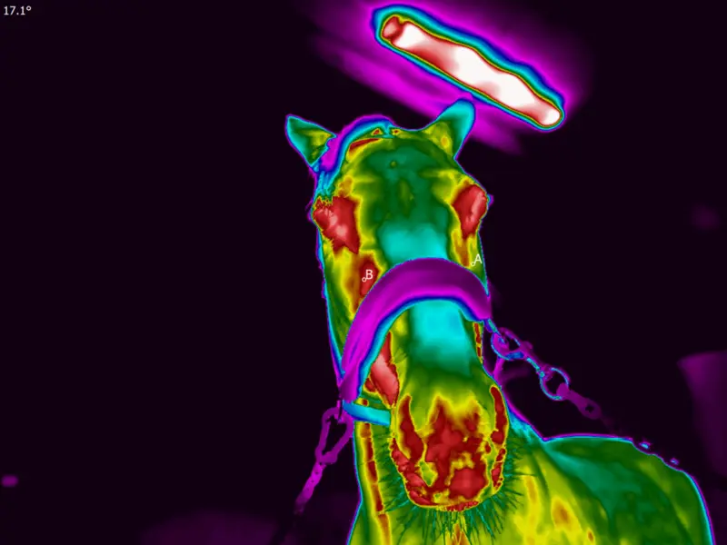
Thermal images showed the problem area to be in the head and neck area. On further examination the horse was found to have an infection in the mouth. The horse had one tooth removed and three teeth were repaired. Thermal imaging also revealed a temperature difference of more than 2°C in the area of the 5-7 vertebrae of the neck. The neck joint problem was treated with an intra-articular injection of cortisone. Bemer therapy was administered as an adjunct treatment.


Neck from below.


Head from below.


Head from the front.
Thermal imaging was a quick and cost-effective way to identify the problem, without causing the horse any pain with examinations. Thermal imaging is a comprehensive, risk-free examination method that allows the veterinarian to quickly and reliably identify potential problem areas for further investigation and treatment.
Discover our new, portable solution. Compact and easy to use, the IRT-384 Tablet allows you to conveniently take and analyze thermal images on a single device - wherever you go.
Learn more and get yours
Thermidas VET
Polttimonkatu 4
33210 Tampere, Finland