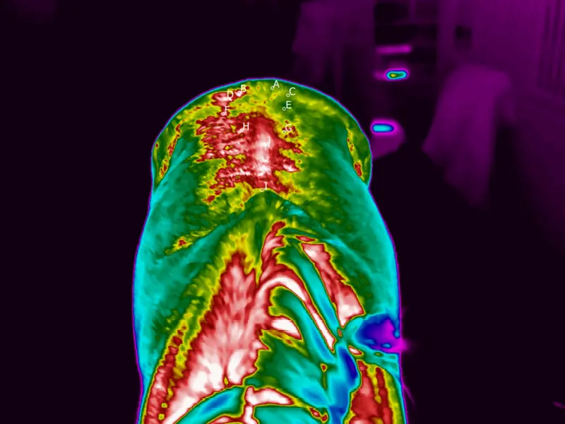
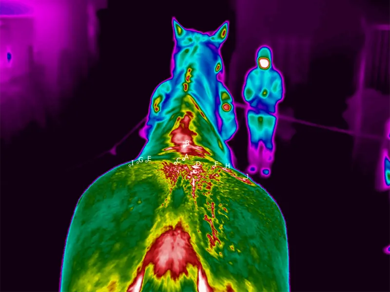
Determining the exact diagnosis of Si joint dysfunction is difficult, due to the often mild chronic symptoms and the challenging location of the joint. The SI joint is difficult to reach because it is located deep in the pelvis and its surrounding bones and muscles prevent its palpation. In general, thermal imaging is a good tool for examining the symmetry of the musculature in the area.
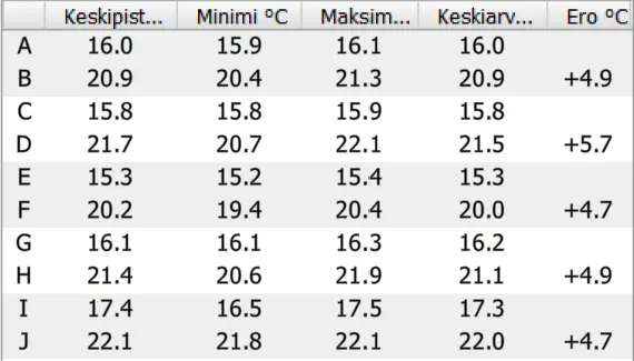
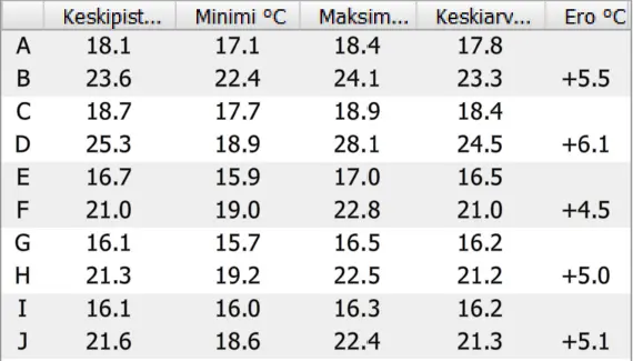
The advantage of thermal imaging is its ease of use, affordability and painlessness for the animal. Thermal images can also be interpreted immediately after imaging, and side differences in temperature can be immediately detected, which when exceeding more than two degrees, can indicate health problems.
Initial situation
A trotter started to act unpredictably and became dangerous for himself and the rider. The horse was examined at a horse clinic, where the focus was mainly on foot care. The response to the treatment was weak and since the horse’s condition seemed serious, euthanizing the horse was considered as one option. The owner contacted our equine expert Mari-Tiina Jääskeläinen, who actively uses thermal imaging in her work.
Findings and treatment
Jääskeläinen took thermal images of the trotter and discovered a thermal anomaly in the region of the SI joint. At the horse clinic, the diagnosis was confirmed as a problem in the SI joint area and a torn ligament. The SI joint was treated with cortisone. Shock wave treatment activated tissue metabolism, restarted the healing process of a prolonged inflammatory reaction and increased the flexibility of scar tissue. The trotter rehabilitated and performed during the competition season as expected.
IRT-384 Tablet
Discover our new, portable solution. The compact and easy-to-use IRT-384 Tablet is a complete solution for taking and analyzing thermal images wherever you are.Learn more and order now